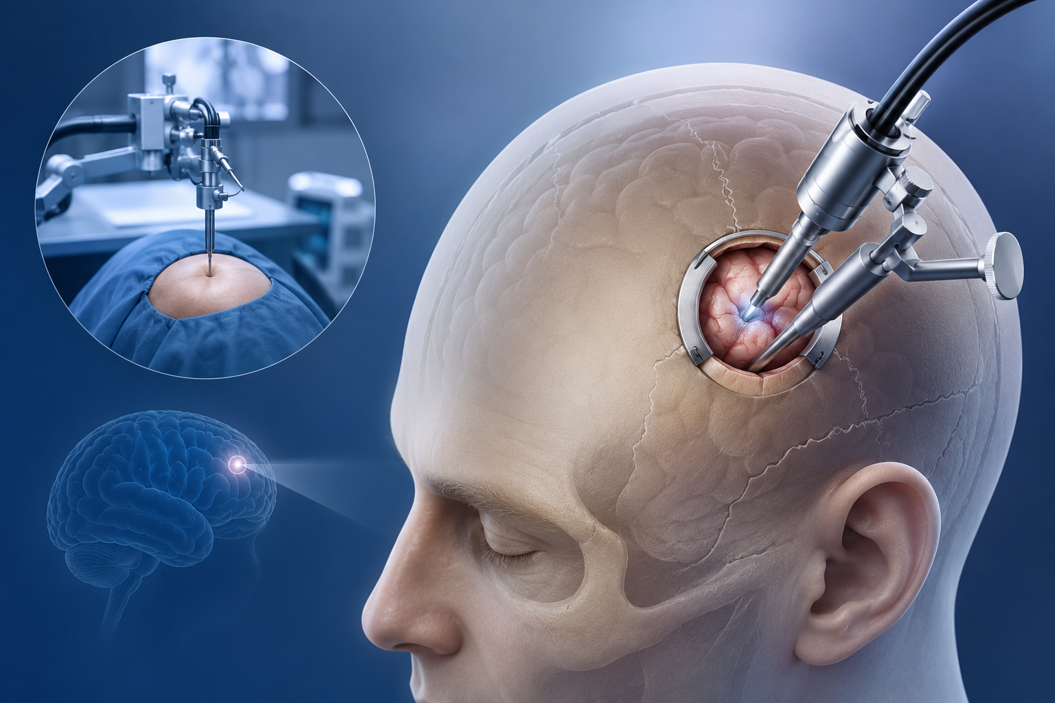
Neuronavigation maps the brain in 3D, allowing for precisely targeted excision through an opening of less than 3cm.
Neurosurgery is no longer characterized by large, traumatic incisions. With the advent of neuronavigation—often described as "GPS for the brain"—we can now reach and remove complex tumors through remarkably small openings, preserving healthy tissue and accelerating patient recovery.
The Power of Neuronavigation
Neuronavigation involves a sophisticated computer system that integrates preoperative MRI or CT scans with the patient's actual anatomy in the operating room. This creates a "live map" that guides the surgeon's instruments with sub-millimeter accuracy. As seen in the navigation screen above, we can visualize the exact boundaries of the tumor and the safest path to reach it before we even begin.
The "Keyhole" Advantage
Traditional craniotomies required large skull removals to provide adequate visibility. Today, navigation allows us to perform "keyhole" surgery. By knowing exactly where the tumor lies, we can create an opening—often less than 3 centimeters in diameter—directly over the target. This results in:
- Minimal Brain Retraction: Less pressure on healthy brain tissue.
- Reduced Blood Loss: Smaller exposure means significantly less bleeding.
- Faster Wound Healing: Smaller incisions heal more quickly and with less discomfort.
- Shorter Hospital Stays: Many patients can return home much sooner than with traditional surgery.
"Precision is our greatest tool. By using navigation to guide our approach, we minimize the impact on the patient while maximizing the surgical outcome. It's about doing the most work through the least amount of disruption." — Dr. Agnetia Vinoth
Advanced Tools for Complex Cases
In addition to navigation, we utilize high-definition exoscopes and neuronavigated ultrasonic aspirators to gently remove tumor tissue. This "gentle surgery" approach is particularly vital for tumors located near critical eloquent areas of the brain that control speech, movement, or vision.
If you or a loved one are facing a brain tumor diagnosis, ask about the possibility of a navigation-guided, minimally invasive approach. Modern neurosurgery is about reaching the target with the utmost respect for the brain's delicate architecture.
Written by Dr. Agnetia Vinoth
Dr. Agnetia Vinoth specialized in advanced neuronavigation and keyhole surgical techniques for complex brain and skull base tumors.
Book a Consultation